Research

Driven by hypothesis, technology and curiosity

One of the major challenges in cell biology is to understand how biochemical reactions at the molecular scale self-organize to generate physiological forms and functions at the cellular and tissue scale. Although genetic and biochemical approaches have been invaluable in identifying key regulatory molecules, these classical approaches rarely have the temporal and spatial resolution required to reveal the molecular mechanisms underlying such self-organization in vivo. Our research aims to address this knowledge gap by developing innovative molecular actuators that can be used to perturb and assay molecular actions in live cells with high temporal and spatial precision. In the past decade, we have applied these tools to studies of innate immune functions such as chemotaxis, phagocytosis and degranulation, as well as subcellular organizations including primary cilia, microtubules and stress granules. Please see below for our recent achievements under this theme!


"Non-catalytic allostery in α-TAT1 by a phospho-switch drives dynamic microtubule acetylation."

Journal of Cell Biology, 2022:221:11:e1-20.


"Location, Location, Location"

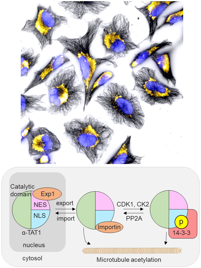
We report an unconventional regulatory mechanism of α-TAT1, a posttranslational modification enzyme that acetylates microtubules. More specifically, we identified a unique signal motif in the intrinsically disordered region of α-TAT1. Multidisciplinary characterization subsequently revealed that this previously uncharacterized motif functions as an intricate molecular switch that integrates upstream kinase signaling to actuate the intracellular localization of α-TAT1, hence achieving dynamic microtubule acetylation. Activity of cellular enzymes is often regulated allosterically, where the interaction between a given enzyme and its substrate is initiated by a third molecule binding to the enzyme to induce conformational change. In contrast, it appears to be the spatial regulation that mainly controls the functional output of α-TAT1. While there are enzymes that translocate between nucleus and cytosol, α-TAT1 is the only enzyme to our knowledge that uses the nucleus as a sequestration hideout.

Technological advance: In parallel to characterizing α-TAT1 regulatory mechanisms, we developed a co-recruitment assay based on chemically inducible dimerization tools to assess protein-protein interactions. This assay is like performing Co-IP assays in living cells, and is quantitative, robust and reliable. Most existing protein-protein interaction assays require purification of proteins and/or lysing cells where procedural concerns need to be taken into consideration for data interpretation. Our co-recruitment assays circumvent this problem. And thanks to the modular design principles, the assay is generalizable to virtually any soluble proteins of your choice. Therefore, we expect its wide application to diverse systems. (Photo credit: Abhijit Deb Roy)


"A molecular trap inside microtubules probes luminal access by soluble proteins."

Nature Chemical Biology, 2021:17:888-895.


"Illuminating the path to cellular bower"

We present the first method to chemically and inducibly trap proteins inside microtubules that enables direct assessment of biomolecular accessibility in living cells. This work overcame previous technical limitations and provides a novel mechanistic insight of how protein regulators and effectors gain access to the site of their action inside the cellular "tunnel".

Microtubules are the fundamental basis for many cellular functions including cell division, cell migration, neuronal plasticity and ciliogenesis. The multi-tasking schemes of microtubule functions is believed to be encoded in a spatio-temporal pattern as well as a combinatorial set of their posttranslational modification status, a concept put forth as the tubulin code. While these modifications mostly occur on the outer surface, microtubules can also be regulated via the inner part, such as via acetylation. As a cellular tunnel, the microtubules uniquely present a lumen that is physically insulated by the cylindrical wall of densely-packed tubulins, with limited connections to teh cytoplasm at their open ends. It is natural to wonder how regulatory and effector proteins of acetylated tubulins reach this secluded site.

The bottleneck of microtubule studies thus far is the limited number of experimental systems. To overcome this challenge, we rationally established a chemically-inducible technique for conditional trapping of a cytosolic protein in the microtubule lumen, with which we subsequently discovered that soluble proteins can enter the lumen via diffusion through openings at the MT ends and sides. Additionally, proteins forming a complex with tubulins can be incorporated to the lumen through the plus ends. We believe that our molecular tool is unique and powerful in probing biomolecular accessibility, with a strong potential in decoding tubulin codes in teh near future, thus appealing ubiquitously to a large audience in cell biology. (Photo credit: Yuta Nihongaki)


"Rational design and implementation of a chemically inducible hetero-trimerization system."

Nature Methods, 2020:17:928-936.


"The Three Musketeers"

We report the first chemically inducible "trimerization" system (CIT), where a small molecule rapidly brings three unique proteins together in living cells, offering unprecedented intracellular operations in addressing new biological questions, harnessing cell functions and behaviors, as well as performing synthetic operations.

Since the development of a chemically inducible dimerization (CID) in the late 90's, a number of CID tools were generated and used to advance life sciences in answering biological questions. In addition, the CID-mediated operation became a foundation for designing optogenetic dimerization tools in recent years. Despite the prevalent trimerization events in nature (death receptors including apoptosis, G proteins initiating signal transduction, etc.), and despite the self-evident utility of CIT, we do not have CIT tools yet in our hands; this is simply because developing CIT technology is extremely challenging.

In this study, our multidisciplinary team constructed a trimerization system by co-opting unique properties of the well-established FKBP/FRB/rapamycin dimerization system, thus rationally avoiding the challenging task of engineering multiple cooperative protein-small molecule interactions de novo. Through computational analysis, we identified split sites for the FKBP or FRB proteins such that the two split protein halves and the cognate protein partner form the three stable protein components of the trimerization system. Live-cell imaging experiments and structural validation with protein crystallography showed that the split-based CIT system undergoes rapid trimerization upon rapamycin addition. Increased capability in spatiotemporal control conferred by CIT allowed us to to demonstrate its potential, for example, in the emerging field of organelle-organelle membrane contact sites. Since CIT molecular tools are modular in design, these first CIT tools pave the way for future generalizable CIT-based approaches that alter function at organelle junctions and ofer higher spatiotemporal control in other biological contexts. This work was highlighted in Nature Chemical Biology, "Three's company". (Photo credit: Helen Wu)


"Intracellular production of hydrogels and synthetic RNA granules by multivalent enhancers."

Nature Materials 2018:17:79-89.


"Making gels in cells"

The physical state of a given molecular assembly in cells is recently proposed to determine its function. To experimentally test this emerging concept is challenging due to limited techniques that can control physical states such as solid, gel and liquid. In the past, researchers have injected hydrogels into cells, only to find their unacceptable cytotoxicity. Noe of the de novo syntheses of hydrogels have seen fruition.

In this study, we rationally developed "iPOLYMER", a strategy for rapid induction of protein-based hydrogel formation inside living cells, by taking advantage of a chemically inducible dimerization paradigm. We then utilized the iPOLYMER technique to probe the effect of physical states of intracellular organizations such as primary cilia and RNA granules. First, we interfered with intraciliary trafficking events by modifying the molecular sieving property at the base of the primary cilia (Nature Chemical Biology 2012). This enabled us to directly study teh role of cilia trafficking without disrupting their delicate structure. Second, we aimed to generate a synthetic mimicry of RNA granules, which are composed of RNAs and proteins. RNA granules are receiving intensive review due to their unique phase-related physical properties as well as their mysterious contribution to biological functions. With iPOLYMER, we successfully reproduced synthetic RNA granules based on non-natural building blocks whose molecular profile perfectly matched with that of physiological stress granules.

To the best of our knowledge, our study is the first successful report of making intracellular gels. We provide a technology for probing biological materials in intact cells, further extend conventional biochemical and biophysical techniques, and offer implications for the progression of diseases caused by protein polymers such as neurodegenerative and prion diseases. This work was highlighted in a press release, "Researchers Report First-Ever "Hydrogels" Made in Living Cells". (Photo credit: Hideki Nakamura)


"Dynamic Remodeling of Membrane Composition Drives Cell Cycle Through Primary Cilia Excision."

Cell 2017:168:264-279.


"Meshing Two Gears"

How the cilia life cycle is linked to the cell cycle remains unknown despite the close relationship between the two events. In this study, we reported a novel and highly dynamic form of organelle disassembly that is precisely controlled by intraciliary regulation of the PI(4,5)P2 membrane lipid. We were the first to comprehensively describe this phenomenon, termed "cilia decapitation," at the cellular and molecular levels. In particular, we showed that the active removal of PI(4,5)P2 from the distal half of cilia membrane allows cilia to form a stable structure by preventing actin polymerization-mediated membrane deformation. Upon stimulation of cell growth, however, primary cilia lose this privilege as PI(4,5)P2 starts to accumulate at the distal half. This dynamic remodeling of membrane lipids triggers a chain of molecular events including actin polymerization which destabilizes membranes and eventually drives cilia decapitation.

We further shed light on the physiological role of cilia decapitation and demonstrated that it not only triggers cilia disassembly but also facilitates cell cycle reentry, thus linking the cilia life cycle to the cellular proliferation cycle.

These discoveries were made possible by a unique combination of different approaches. Besides live-cell and super-resolution fluorescence imaging, revealing sub-micron and sub-minute resolution of this dynamic process, newly developed genetically encoded molecular actuators allowed us to achieve molecular perturbation specifically inside the primary cilia. Previously, perturbation of actin polymerization was limited to pharmacological interventions, the effects of which are not confined to the primary cilia. In summary, our study is conceptually and technologically novel, offering far-reaching implications for understanding ciliopathies. This work was highlighted in a press release, "Some Cells Need a 'Haircut' Before Duplicating", and the PKD News Blog titled "Study Shows How and Why Hairlike Structures on Cells are Lost". (Photo credit: Siew Cheng Phua)