Injury Models of SCI
The Contusion Model
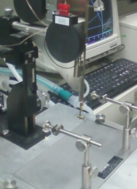
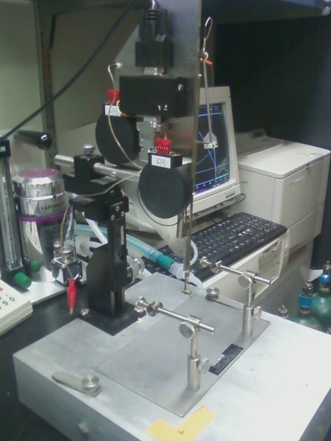
A laminectomy, or the removal of the lamina of the vertebral column, is a secure and simple way to expose the spinal cord. After setting the computer to record the dynamics of the impact trajectory, a pin suspending the impact shaft is released and allowed to descend by gravity to hit the cord. The contusion impact velocity and compression is monitored to guarantee consistency.
Unilateral Injuries


In addition to a simple midline weight drop, we are also able to induce a spinal cord injury to only one half of the spinal cord, known as a unilateral injury. These models are ideal for evaluating specific motor pathways, such as the corticospinal (CST) and rubrospinal (RST) tracts, and for assessing the efficacy of rehabilitative therapies. For example, many studies have shown that the motor tracts are able to sprout new lateral axons into the undamaged region of the spinal cord after a unilateral injury.
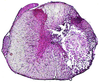
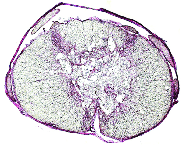
The Compression Model
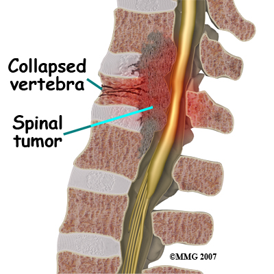
Compression injuries can be achieved in animal models by several ways: applying pressure to the exposed spinal cord using a clip, injecting cancer cells that grow into a tumor in 3 to 4 weeks (2), or inflating at an epidural balloon under the vertebra (3).
Experimental Autoimmune Encephalomyelitis
Experimental autoimmune encephalomyelitis (EAE) is a rodent model of an inflammatory demyelinating disease of the central nervous system. In one version of EAE, purified myelin oligodendrocyte glycoprotein (MOG) is used as an immunogen in combination with an adjuvant of either CFA or IFA to induce a cellular and humoral immune response against the myelin. The MOG is expressed on the external surface of oligodendrocytes and is crucial in the process myelinating of axons. It is also a target of the immune system in demyelinating diseases such as multiple sclerosis (MS).
MOG IFA immunized rats can be induced to have focal spinal cord disease by administering inflammatory factors directly into the spinal cord. This focal EAE model, analogous to the human paralyzing disorder transverse myelitis (TM), is believed to be a valuable model to study the neurological manifestations of a single inflammatory/demyelinating lesion. Using this model, researchers have found extensive axonal degeneration following a single inflammatory lesion.
- Image and compression model information courtesy of utg1.uptontechnologygroup.com
- Ushio Y, et al. Experimental spinal cord compression by epidural neoplasm. Neurology. 1977 May;27(5):422-9.
- Guizar-Sahagun G, et al. New approach for graded compression spinal cord injuries in Rhesus macaque: method feasibility and preliminary observations. J Med Primatol. 2011 Jul 7. doi: 10.1111/j.1600-0684.2011.00483.x.